Suppose that in an office, several letters are being issued in each and every member’s name separately from a single source for any purpose. Now, only those letters which bear a correct postal code or address of the receiver will reach its target destination. But those with any error in the postal code or address will not reach the target receiver. Likewise, all proteins are synthesized on ribosomes in the cytosol regardless of its target site of action. So, naturally a question will arise – How all proteins are directed to their individual final cellular destinations appropriately? Because proteins don’t know where to go and work. Does there exist any sort of postal code even in proteins that are playing the pivotal role? So, let’s understand this complex and fascinating process, particularly for eukaryotic cells.
THE SIGNAL SEQUENCE
In eukaryotic cells, all proteins start their synthesis in the cytosol but not all of them complete the job here. It depends on the primary cellular destination of the proteins. Proteins destined for secretion, integration in the plasma membrane or lysosomal inclusion, are all directed to the endoplasmic reticulum. Some proteins are destined for the nucleus, the mitochondria and the chloroplasts. Remaining ones which are destined for cytosol simply remain where they are synthesized.
In 1970, scientist Gunter Blobel and his colleagues first postulated that this targeting of proteins to their cellular destinations entirely depends on a short stretch of amino acids mostly in the N-terminal of the nascent polypeptide which is called Signal Sequence. It directs a protein to its appropriate primary cellular location and in most of the cases, is cleaved after its job is done. Thus, it acts basically as a postal code of the proteins. It is confirmed by fusing the signal sequence of one protein in a second one and showing that the second protein is heading towards the destination of the first protein. They vary in length but have the following listed features in common:
- About 10 to 15 hydrophobic amino acid residues.
- One or more positively charged residues at the beginning.
- A relatively polar short sequence of amino acids at the carboxyl terminus.
- A cleavage site.
Let’s focus on the first category of targeting i.e. Endoplasmic Reticulum. Shall we?
JOURNEY TOWARDS ENDOPLASMIC RETICULUM
The best characterized targeting system occurs in the Endoplasmic Reticulum (ER). Most proteins: membrane, secretory and lysosomal, have a kind of signal sequence in its amino terminal which targets them for localization in the ER.
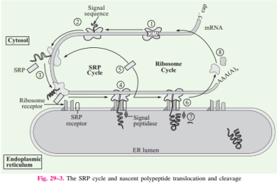
- Protein synthesis starts in free ribosomes of the cytosol. As mentioned before, due to the presence of signal sequence in the amino terminal, it is synthesized first.
- When it emerges out of the ribosome, the signal sequence along with ribosome is recognized and bound by a Signal Recognition Particle (SRP).
- SRP halts elongation of the polypeptide by restricting the passage of translation factors to the A site of the ribosome by binding to GTP.
- Then, the GTP bound SRP directs the ribosome and the incomplete polypeptide to the SRP receptors present on ER.
- The nascent polypeptide then is channelized to peptide translocation complex in the ER where it interacts with the ribosomes bound to ER.
- SRP dissociates by GTP hydrolysis and translation continues now.
- Finally, when the complete protein is synthesized, the signal sequence is cleaved off by signal peptidases within the ER.
Post Translational Modification In Endoplasmic Reticulum And Golgi Apparatus
The signal peptide directs the proteins to the ER. In the ER, various protein folding, disulphide bond formation and other post translation modifications begin. Glycosylation is an important modification which proteins undergo in ER to increase the proteomic diversity, wherein various carbohydrate moieties are attached to the proteins. Glycosylation helps in targeting proteins to the correct destination, cell matrix adhesion and other signal transduction pathways. Our blood group antigens on RBCs are differentiated by the sugar residues it contain.
D-Glucose, D Galactose, D-Mannose, L-Fucose, N-Acetyl Glucosamine, N-Acetyl Galactosamine etc. are common carbohydrate moieties that participate in glycosylation. Most of the glycosylations which occur are N-linked (i.e. sugar moieties attached to amino group of asparagine) or O-linked (i.e. sugar moieties attached to hydroxyl group of serine or threonine). Besides that, additional modifications occur in the Golgi Apparatus before finally sending them to their final cellular destinations i.e. either cell or organelle membrane, extracellular matrix or lysosomes.
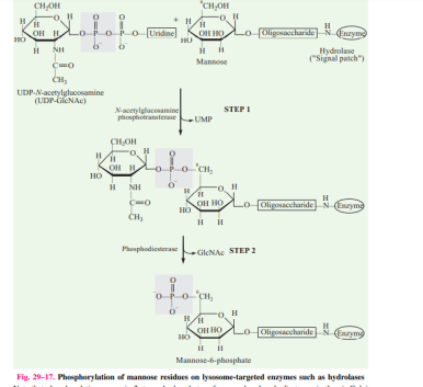
Mannose-6-phosphate: The Lysosomal Code:
In the Golgi, after the proteins have been glycosylated, their final destination journey starts. In proteins destined for lysosomes, a mannose residue on the surface is phosphorylated forming mannose 6-phosphate, which is the structural signal.
THE FANCY STORY OF THE MEMBRANE PROTEINS
Only signal peptide sequence is not enough for proteins to reach their final destination. It needs additional sequences which will guide them towards their destination from the ER or Golgi. Membrane proteins additionally contain some sequences in the middle which are called topogenic sequences (Stop Transfer Sequence and Signal Anchor Sequence). These determine the protein’s orientation in the cell membrane or membranes of ER, Golgi etc. The most important of them is called Signal Anchor Sequence.
It is a 22-25 long hydrophobic amino acid chain which predominantly forms alpha helix with 3 positively charged residues either at its left or right. This determines their orientation in the ER membrane. There are three scenarios:
a. Positively charged amino acids located on N-terminal of hydrophobic sequence: protein will be oriented transmembrane with its N terminus into the cytosol and C terminus into the lumen of ER as positive charges have to stay cytosolic.
b. Positively charged amino acids are located on C-terminal of hydrophobic sequence: protein will be oriented transmembrane with its N terminus into the lumen and C terminus into the cytosol.
c. For multipass protein, the scenario becomes complex because here, the position of N terminus will depend on orientation of first signal sequence. The position of C terminus will depend on the total number of stop transfer and signal anchor sequences. If it is even, then C terminus will end in same side of N terminus. But if it is odd, the C terminus will end in the opposite side.

Most proteins leave the ER, transported in vesicles to the Golgi and from there they are either secreted, or travel to cell membrane, or bound for lysosomes. Likewise for membrane proteins, secretory proteins must also contain some additional conserved sequences for their transport towards extracellular matrix.
CONCLUSION AND QUESTIONS:
So far we have seen the role of signal peptide sequences and other additional sequences within a protein which are the sole determinants of a protein’s final destination. This article focuses on proteins which head towards ER i.e. a majority fraction of membrane, secretory and lysosomal proteins. In addition to that there are proteins which travel towards mitochondria, nucleus, chloroplast etc. We have seen that every time signal peptides are cleaved after its job is done. But there are some proteins mostly nuclear proteins whose signal sequences are not cleaved and also some others which do not have any kind of signal sequence from the beginning. So the story is not over yet. I will conclude here with some questions which need to be addressed in this regard:
- What signal sequence(s) is/are responsible for selectively phosphorylating mannose residues on the lysosomal proteins? Why is there a bias for phosphorylation on mannose especially?
- Is signal peptide mRNA transcribed/translated along with the protein coding region or is it transcribed/translated separately from a different region of DNA and then gets combined with the protein part?
- What is the exact reason of such conserved features of signal sequence?
- Which additional sequences or mechanisms determine whether a membrane protein will be inserted in cell membrane or an organelle’s membrane?
REFERENCES:
i) Lehninger’s Principle of Biochemistry, 7th edition
ii) Cell Biology 04: The Secretory Pathway- Harvard University Cell Biology Lecture Notes